
July 16, 2017, updated May 2019
For many decades people have claimed that the human eye is poorly and inefficiently designed, mostly by comparing it with the eye of cephalopods (octopi and squid). Though for the past 30 odd years this has been known to be false, yet this falsehood persists and continues to promulgate to this very day. In this article I will discuss the features of the human and cephalopod eye, show the reasons for the design of the human eye, and indicate why its design is optimal for its purpose.
I first studied the eye in the late 1980s when I spend two years as a teaching assistant for Dr. Werner K Adrian who taught the second year colour vision course in the Optometry Dept. at the University of Waterloo.
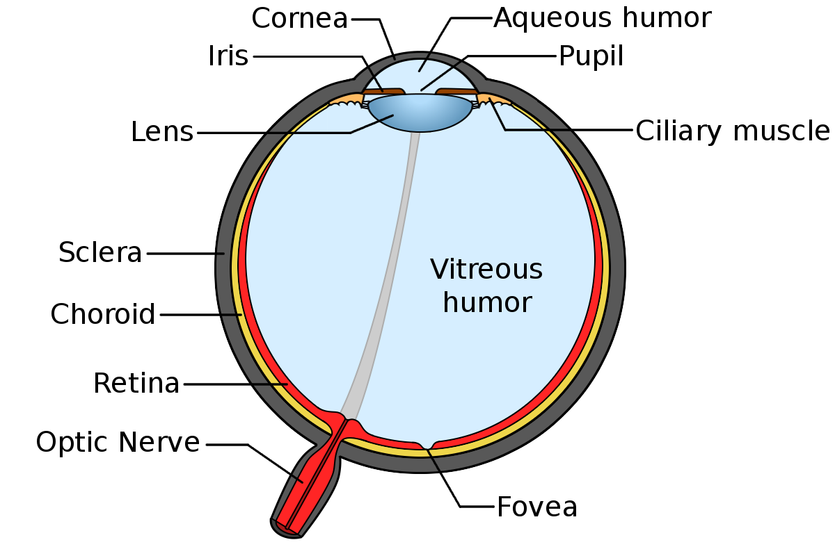
1. The Human Eye – general overview
It’s helpful if you have a general knowledge of how the eye works, but there is not time to go into much detail here. Later on I’ll be focusing on the retina and optic nerve in more detail.
The Minimum Requirements for vision:
• very transparent cornea
• very transparent lens
• very transparent vitreous & aqueous humor
• light sensitive cells (we have two types with 4 colour sensitivities)
• photoreceptors connected to nerve cells & optic nerve
• curved cornea for main focus
• adjustable lens for fine focus
• lens focused to some useful distance
• lens and humours must have different indices of refraction
• eyelids and tearducts for protection and cleaning
• process of removing dead cells so that they don’t clog up the eye
 A
A
Extra features of the Human Eye:
• recessed eyes (protected by bones)
• colour vision
• vision over huge range of light intensities
• pigmented iris to block light except through pupil
• moveable iris that controls amount of light
• eyebrows to keep water out of eye
• eyelashes to protect against dust
• binocular vision
• automatic feedback (focusing, iris, tracking)
• saccade movements (to prevent photoreceptor fatigue)
• excellent focus at fovea — able to read text
• macula lutea (extra UV protection on fovea)
• colored iris – beauty
2. Cephalopod Eyes
The eyes of squid and octopi are quite similar to ours in their general construction. Each is a single camera-like structure with basically the same components.
There are some significant differences between the two types of eyes. Cephalopod eyes contain only one type of photoreceptor so they cannot detect colours, only shades of gray. (The colours that could be seen are limited because not as much red light penetrates to the depths where they live.) These receptors are called rhabdoms (and are also in insect eyes where they are part of a structure called an ommatidium) and are roughly equivalent to the rods in vertebrate eyes.
Unlike our eyes, these receptors can detect polarized light (as they also do in insect eyes). In the ocean there is a 10-fold drop in brightness with every 75 m depth increase1, so most cephalopods are not exposed to bright light and cannot see in bright light. (The octopi that live in shallow waters probably have eyes that are adapted to higher light conditions.) Because cephalopods only have one type of photoreceptor, they cannot see as great a range of light intensities as we can with the combination of our rods and cones. The brightest object that we can see is 1000 billion times brighter than the darkest one.2, 3 Cephalopods don’t change the shape of the lens to focus, but instead move the lens forwards or backwards in the eye to adjust focus. They have very little spherical aberration4 (probably due to gradient lens such as humans have). The healthy human eye can focus from 6.5 cm to infinity which is a range of 15 diopters. Cephalopods can only change the accommodation of their eyes by 5 diopters5.
One of the most interesting features of cephalopod eyes are their weird shaped pupils. Not only are these pupils off-axis, but they are shaped as a U, a W, or a dumbbell. This means that they would always have blurry vision and reduced visual acuity – images would never be in clear focus on the retina. What is the reason for this? Well …
While octopi cannot see in colour, they can change the colour of their skin to match their surroundings. How is this possible? Their eyes have a significant amount of chromatic aberration and their strangely-shaped pupils work to maximize this effect. Recently, researchers have deduced that this allows them to selectively focus different colours on their retina at one time (e.g. they may put yellow objects in focus and make blue ones blurry)6, 7. This unusual mechanism of detecting colour differences requires extra visual processing to analyze it, but it seems to work for them.
Most cephalopods are visual hunters, so it is probably this colour detection combined with binocular vision that enables them to see their prey and determine how far away it is in spite of their blurry vision.
Thus, the human eye is superior to cephalopod eyes in the following areas. We have …
• clearly focused images
• much better visual acuity (an octopus eye would never be able to read text)
• colour vision (all colours at once)
• much greater range of focusing
• much greater range of light intensities
Conclusion #1: The human eye is indisputably superior to the cephalopod eye.
No sane person would actually want to trade his human eye for the cephalopod eye (if it were indeed possible to do this).
Are there ways in which the cephalopod eye is superior to human eyes?
The oft repeated maxim is that octopus eyes are better than human eyes because unlike theirs, our retinas are inverted and have a blind spot. This is posited as a very poor design compared with our cephalopod friends. Let’s examine this more closely.
3. The Cephalopod Retina
It has proven to be impossible to find detailed diagrams of the cephalopod retina showing all of its layers. As seen in the diagrams below , they just show the photoreceptors and the nerve cells in a loose bundle of ganglia heading to the brain.
 B
B 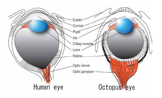 C
C
It is possible that there is a retinal pigment epithelium behind the retina, but the diagrams do not show this. Nor does it show a choroid layer. The sole significant and oft-repeated feature of the cephalopod retina is that light coming into the eye hits the photoreceptors in the retina immediately and directly without having to pass through layers of nerve cells and blood vessels.
The Human Retina:
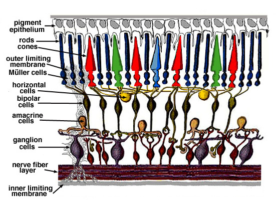
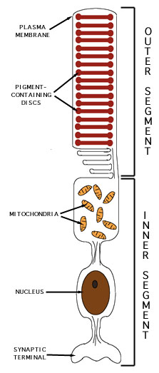
The diagram below clearly shows the various layers of the human retina. Light comes into the eye from the bottom of the diagram and moves upwards until it hits the outer segments of the rods and cones where it is detected and changed into nerve signals. Not shown is a spider-web of blood vessels on top of the nerve fiber layer; yes, light has to go past blood vessels too.
 D
D
So, a terrible design? Not exactly. It’s really a very clever design working within and overcoming some specific design constraints that are signficantly different from the cephalopod eye. Deciding what is a good or bad design is somewhat subjective. Those who claim that the human eye is poorly designed end up saying that evolution has added some very clever and ingenious mechanisms to compensate for the initial poor design. However, this could equally well be interpreted as a biochemical engineer looking at how to design a highly efficient eye and realizing that the retina has to be inverted for it to work (for reasons explained below). The various techniques used to get light to the outer segments are thus not kludges to overcome a stupid design decision, but intrinsic parts of a very complex and effective design. The main constraint is that it is absolutely essential that the outer segments of the photoreceptors be embedded in the retinal pigment epithelium (RPE) in order for them to function and to reduce light scattering in the eye.
The outer segments of the rods and cones contain discs where the light sensitive opsin molecules are. This is the part of the photoreceptor cell that detects light and then sends a signal across the synapse to the nerve cell. The RPE performs at least seven distinct functions8. I’ll discuss 3 of them here.
ref. E
First of all the RPE is highly pigmented so that it absorbs light. This prevents any photons that don’t get absorbed in the outer segments from scattering and blurring the image. (For animals that have a tapetum, the RPE is transparent and the tapetum behind it reflects the light back through the rods or cones, giving them a second chance to detect the photons – and making their eyes appear glowing green at night.)
Secondly, the outer disks wear out quite rapidly. They are formed near the middle of the photoreceptor and move up to the tip over 11 days, where they are pinched off and discarded. The RPE digests these worn out disks (phagocytosis) so that they don’t clog up the retina and prevent the photoreceptors from getting the nutrients it needs. When this process malfunctions, the retina degenerates.
Thirdly, the RPE and the choroid layer behind it provide oxygen, nutrients and necessary chemicals for the photoreceptors to function. These include glucose, fatty acids, and retinal (the molecule, not the adjective). When light strikes 11-cis-retinal (in the disks in the rods and cones) it changes to all-trans-retinal which then triggers changes in the opsins. The outer segments do not regenerate the retinal back into the cis- form once it has been changed by light. Instead the retinal is pumped out to the surrounding RPE where it is regenerated and pumped back into the outer segments – ready for action again. This is probably so that the outer segments don’t get cluttered up with extra chemical machinery; anything not absolutely necessary is outsourced to the RPE.
So, why do the rhabdoms in the cephalopod eye work without being embedded in the RPE? Rhabdoms are a far simpler structure that is found in insect eyes as well. The Encyclopaedia Britannica says9 “In most invertebrate photoreceptors the structure is different [from rods and cones], with the photopigment borne on regularly arranged microvilli, fingerlike projections with a diameter of about 0.1 μm. This photoreceptor structure is known as a rhabdom. The photopigment packing is less dense in rhabdoms than in vertebrate disks.” Rhabdoms also frequently “contain granules of dark pigment that move toward the rhabdom in response to light. They act as a type of pupil, protecting the rhabdom in bright conditions by absorbing light.” Note that rhabdoms are simpler, are less chemically active (with less rhodopsin), and probably less sensitive than our photoreceptors.
What about the blood vessels? They provide nutrients to and remove waste from the inner retinal layers; the RPE and underlying choroid to this for the outer layers. Note that the choroid supplies about 75% of the blood to the retina and the retinal vasculature only 25%.10 If one had to remove one of these two sources of nutrients, it’s the blood vessels that would go because the RPE is so essential. This, in fact, is what happens in the avian eye. They have no retinal blood vessels at all, but have a special organ called the pecten oculi that helps supply oxygen and nutrients. Their eyes are significantly smaller than mammal eyes so oxygen, glucose and CO2 don’t have to diffuse as far.
The blood vessels perform another vital function. The cells which have the highest metabolic activity in the body are the cone cells. Professor of Opthamoloty, Alfredo A. Sadun, says “It may be counterintuitive, but neurons, with no moving parts, have a lot of mitochondria. Neurons are very high maintenance because of the action potentials and the need to re-establish the sodium and potassium gradients after every action potential. But the greatest concentration of mitochondria in the body is in the retina. Specifically, rods and cones have a specialized part, called the inner segment, which is stacked with nothing but mitochondria. Each rod has several hundred mitochondria jammed into this portion which is closest to the outer segment where light creates signal. The cones have even more mitochondria than the rods. There is no tissue in the body that has a greater concentration of mitochondria. Vision does not come cheap.”23
This quote explains the necessity of extra blood vessels in the retina to nourish the neurons, instead of just relying on the choroid for this. Now, all of this metabolic activity in the outer segments of the photoreceptors generates a lot of heat. The inverted retina allows the blood supply of the choroid to remove this heat. If the retina was not inverted, the heat would have nowhere to go. The vitreous humour is gel-like and does not have convection currents that can help cool the retina. The whole eye would eventually heat up and begin to be damaged by the build up of heat.
Conclusion #2: The human retina must be inverted if it is to function at all.
All of this has established the importance of having an inverted retina. How then do we have clear vision in spite of all of the layers of the retina that the light must go through before it is detected?
4. Compensating for our inverted retina
How does light get to the outer segments undistorted? There are a number of features that are in play here.
- The neurons in the retina are transparent and have to be stained in order to see them under a microscope.11
- The rods and cones act like tiny fibre optics transmitting any light received at the near end to the far end.
- Recently, researchers found that the Müller glial cells that provide scaffolding and other functions for the retina also act as fiber optic guides for light12, 13 . They take the light past all of the intervening layers and direct it to the rod and cone outer segments. You can see one of these cells in the diagram of the retina above.
- Not only do they act as living wave guides, Müller cells also separate the red and green light from blue and direct the former to surrounding cones in order to “to improve day vision with minimal effect upon night vision”14
- What about the blood vessels that crisscross the retina?
- The capillaries are embedded in the layers of the retina15 so their effect is counteracted by the Muller optical fibres that take the light past all of these obstacles.
- The larger arterioles and venules seem to lie on top of the inner surface of the retina, so they would cast a shadow onto the photoreceptors. Since the blood vessels do not move with respect to the retina, the image is static. The brain ignores stationary images (which is why our eyes are continually moving) so we don’t see these shadows. The arteries and veins must still reduce our visual acuity to some extent, but they comprise less than 10% of the surface of the retina.
The optic nerve and blind spot
Because the layers of nerve cells have to send their signals out of the eye to the brain, there must be an optic nerve that pierces the retina. This creates a blind spot (aka optic disk) in each eye, a place where there are no photoreceptors, only nerve cells. This spot is approximately 6° wide. Does this blind spot cause all sorts of problems for us? No, actually it doesn’t. The blind spot is off to one side and the blind spots in our two eyes never overlap. Our eyes are continuously moving so the rest of the retina detects what’s in that area, and the brain builds a complete image of our surroundings. One never hears someone say, “I walked into the door or stubbed my toe because it was in my blind spot”. I can’t even imagine how one could intentionally set it up to injure ones’ finger or toe because of the blind spot. It would take a lot of ingenuity and careful planning to do this.
Since there can be no photoreceptors where the optic nerve is, various other things that might also interfere with vision are placed there. The retinal artery and vein enter and exit the eyeball through the blind spot. The avian pecten oculi is located there. And the hyaloid canal to the lens originates there (it is the grey line going through the eye in the first diagram). The hyaloid canal is used to take an artery to the lens in the fetus when the lens is forming and growing. Once the lens is the correct size, the cells in it become essentially dormant and are almost dead. The hyaloid artery is no longer needed and regresses. (There is some suggestion that the hyaloid canal also allows the eye to compensate for volume changes in the lens with changing focus. However, more recent studies have found that the volume of the lens is nearly constant no matter what the accommodation is. Also the hyaloid canal is filled with liquid, which being incompressible, would not be able to compensate for volume changes in the lens.)
The part of the eye that has the best focus is called the fovea. The retina at the fovea is very thin, with at most one layer of neurons over it. There are also no blood vessels at all in the fovea. The fovea is packed as tightly as possible with cone cells, and each cone cell is thinner than cone cells elsewhere in the retina. All of this gives excellent resolution at the fovea and enables us to read very small text. Many people with good eyesight can read the microprinting that makes up the signature line of cheques even though the letters are only about 1/3 mm (less than 1/64 inch). Our fovea has amazingly excellent vision.
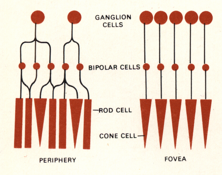
Before we finish with the retina, we need to note one more thing. The retina does a lot of image processing before the signal is sent to the brain.10 The “photoshopping” that goes on in the neural layers of the retina primarily does three things: data compression, edge enhancement, and maintaining colour constancy in different light conditions.16 The main reason for this is to reduce the number of ganglia that are needed to transmit a signal to the brain via the optic nerve. This keeps the optic nerve down to a manageable size yet still produces a good image. Note that in the fovea, in order to have the best vision possible, every single cone has its own ganglion cell that transmits the signal from the cone directly to the brain. The diagram below illustrating this is from it the TimeLife book “Light and Vision” from 196617. The data compression that the retina performs is not a simple fixed process: it adapts to light levels. For example, assume that in some part of the retina five rods and cones connect to one ganglion (after various processing in the intermediate layers). When the light levels decrease, neural network in the retina reprograms itself to group a larger number of photoreceptors together. The retina makes the tradeoff of reducing visual acuity for the sake of being able to detect lower levels of light.18 (The image processing functions of the retina were discovered sometime before 1988 when I learned about them from Dr. Adrian.)
 Is it detrimental to lose so much information from the image before it is processed in the visual cortex at the back of the brain? No, not at all. Obviously, the brain does not store all parts of an image that it sees. It is important to realize that the retina and optic nerve are actually part of the brain (as is the olfactory nerve); they are not part of the peripheral nervous system originating in the brain stem or spinal cord. So it’s not correct to say that the optic nerve transmits the image from the retina to the brain. Rather, the neurons in the retinal part of the brain begin the image processing that the visual cortex would otherwise have to do later on. This is ingenious: at some point the image has to be processed and analyzed. Doing the initial part of it in the retina reduces the amount of data that needs to be sent to the rest of the brain without causing any diminishing of vision; thus, the optic nerve can be reduced in size to something that does not affect our vision in any way.
Conclusion #3: Neither the blind spot nor the inner layers of the retina cause any significant or noticeable problems with our vision.
5. The Promulgation of Errors
So why does this myth of the problematic inverted retina persist?
Well, on the one hand, there are a lot of people who just repeat what they hear. This happens with all subjects and nowadays with the internet and social media everyone thinks that he or she is an expert.
On the other hand, there are people who have a vested interest in believing that the human eye is a poor design and so intentionally perpetuate this urban legend. I’m sure that there are innumerable examples of this since this myth has been around for such a long time; however, I’m just going to discuss the writings of two biologists that I came across who say such ludicrous things like “Indeed, if engineers were to build an eye with the flaws of our own, they would probably be fired.”19 (Note that there are other scientists who recognize the clever design of the eye: “Having the photoreceptors at the back of the retina is not a design constraint, it is a design feature. The idea that the vertebrate eye, like a traditional front-illuminated camera, might have been improved somehow if it had only been able to orient its wiring behind the photoreceptor layer, like a cephalopod, is folly.”14b)
As far as I know, our eyes are still far better than any camera that we can design. Someone actually did the calculations (in 2015) to show that building a camera that could do everything that our eye does would cost $35 million dollars.21 $35 million dollars for one eyeball! As cameras have improved over the years since that article was written, they are catching up to the eye and are getting to the point where one camera can match most of its abilities. Digital cameras can now even cover the huge range of light intensities that the eye can see — a range of 1000 billion times (12 orders of magnitude).22
It should be noted that all of the best eyes in the animal kingdom have inverted retinas. An eagle is said to be able to see a rabbit 3km away. This would be impossible with a non-inverted retina. Furthermore, none of the detractors of the inverted retina have been able to come up with a complete fully developed eye that does everything that ours does and that avoids all of the problems that occur when you put the retina the correct way. They assume that that flipping the retina around and getting it to work as so simple that a two year old could do it.
The first example is Prof. Nathan H. Lents, a molecular biologist, who wrote about the ostensible appalling design of the eye on his blog in 201520. First he claims that the human eye is poorly designed because it is subject to all sorts of diseases and malfunctions. This is basically like saying that because some part of your body will eventually wear out or can be infected, it is not well designed. Secondly, he points out that there are other animal eyes that can do specific things better than ours, such as seeing in the dark, detecting polarized light, seeing into the UV range, hawks can see small creatures from much greater distances than we can. The problem with this is that there is no one eye that can do absolutely everything AND all of these “better” eyes also have inverted retinas. Our eyes are very very good for the purposes that we need them for in every aspect of everyday life. Finally, he brings up the “backwards” retina and blind spot (but bizarrely, the image shown in his blog is of the fovea, not the blind spot!). You don’t have to be in Mensa to do a tiny bit of research to learn that the inverted retina and blind spot are not actually problems. Professor Lents has an agenda: he wants to show that the eye is rife with problems and that evolution did not do a good job of designing the human eye. As he mentions in his conclusion, this is specifically for the purpose of arguing against Intelligent Design.
A more egregious example is an article in Scientific American in 2011 by a Dr. Trevor Lamb.19 Dr. Lamb is a professor of visual neuroscience who actually studies the eye for a living and yet here he deliberately deceives us about the design of the eye:
For all the ingenious features evolution built into the vertebrate eye, there are a number of decidedly inelegant traits. For instance, the retina is inside out, so light has to pass through the whole thickness of the retina–through the intervening nerve fibers and cell bodies that scatter the light and degrade image quality–before reaching the light-sensitive photoreceptors. Blood vessels also line the inner surface of the retina, casting unwanted shadows onto the photoreceptor layer. The retina has a blind spot where the nerve fibers that run across its surface congregate before tunneling out through the retina to emerge behind it as the optic nerve. The list goes on and on.
This backwards retina “problem” has been answered numerous times over the past decade. As I’ve shown, it is not a mistake, but rather an advantage as it allows for much more active photoreceptors. The most chemically active parts of the retina are in direct contact with the RPE and the blood supply of the choroids. The transparent nerve cells do not degrade our vision. As for the blind spot, I’ve shown that it doesn’t affect our activities at all. The blood vessels casting shadows in our peripheral vision may or may not interfere with our vision. I can’t find any research that addresses this and quantifies any reduction of our vision.
Lamb continues
“Although camera-style eyes provide a wide field of view (typically of around 180 degrees), in practice our brain can sample only a fraction of the available information at any given time because of the limited number of nerve fibers linking our eye to our brain.”
The author was just complaining that we have a blind spot, but here he now says that it should be bigger! As any optometry student knows, this limited number of nerve fibers means that our optic nerve and consequent blind spot is limited to a reasonable size. We do have dedicated nerve fibres right where we need them – in the fovea. The retina does a lot of signal processing specifically so that the brain doesn’t have to do it. Again, this sort of information is known to any optometrist. One would expect a Ph.D. to have the basic background needed in his field as well as to present the facts objectively with integrity.
Why is Lamb misrepresenting the facts? In his article, The Evolution of the Eye, he says “the eye, far from being a perfectly engineered piece of machinery, exhibits a number of major flaws—these flaws are the scars of evolution.” Here, again, is an example of someone who pushes an agenda so strongly that he distorts the facts in order to support his argument.
I’ve demonstrated here that the eye is superbly designed. Furthermore, I don’t believe that having a well designed eye deals a death blow to evolution as there are plenty of other superb designs in nature and evolution copes with them just fine. This article is the result of about a week of research. Presumably Scientific American and a professor of visual perception could do their due diligence equally well.
References
All online sources were accessed during the week of July 6, 2017.
I have added a ★ to references that are worth reading for more information.
Diagrams
A. Original source of the diagram is are unknown.
B. Diagram from https://thehumanevolutionblog.com/2015/01/12/the-poor-design-of-the-human-eye/
C. Diagram from https://en.wikipedia.org/wiki/Cephalopod_eye
D. Original source of the diagrams used are unknown. They are found widely dispersed on the internet
E. Diagram from: http://www.chemistry.wustl.edu/~edudev/LabTutorials/Vision/Vision.html
Sources
1. Chung, Wen-Sung, and N. Justin Marshall. “Complex Visual Adaptations in Squid for Specific Tasks in Different Environments.” Frontiers in Physiology. February 24, 2017. Retrieved from https://www.ncbi.nlm.nih.gov/pmc/articles/PMC5323406
2. Schubert, E. F. (2006) Light Emitting Diodes, Second edition. Cambridge University Press. (p.276) Retrieved from https://www.ecse.rpi.edu/~schubert/Light-Emitting-Diodes-dot-org/Sample-Chapter.pdf
3. “Adaptation (eye).” Wikipedia. May 15, 2017. Retrieved from https://en.wikipedia.org/wiki/Adaptation_(eye)
4. Sivak, J. G. (1991) “Shape and focal properties of the cephalopod ocular lens”
Canadian Journal of Zoology, 69: 2501-2506.
Retrieved from http://www.nrcresearchpress.com/doi/pdf/10.1139/z91-354
5. Schaeffel, Frank., Murphy, Christopher J., Howland, Howard C. (1999)
Accommodation in the Cuttlefish (Sepia Officinalis) The Journal of Experimental Biology, 202, 3127–3134
Retrieved from http://jeb.biologists.org/content/jexbio/202/22/3127.full.pdf
6. Sanders, Robert. “Weird pupils let octopuses see their colorful gardens.” Berkeley News. July 08, 2016. Retrieved July 16, 2017, from http://news.berkeley.edu/2016/07/05/weird-pupils-let-octopuses-see-their-colorful-gardens ★
7. Stubbs, Alexander L., and Stubbs, Christopher W. (2016) “Spectral discrimination in color blind animals via chromatic aberration and pupil shape.” Proc. Natl Acad. Sci. USA 113, 8206–8211. (doi:10.1073/pnas.1524578113)
Retrieved from http://www.pnas.org/content/113/29/8206.full.pdf ★
8. Strauss, Olaf. “The retinal pigment epithelium.” Webvision. Moran Eye Center, 2011. Retrieved from http://webvision.med.utah.edu/book/part-ii-anatomy-and-physiology-of-the-retina/the-retinal-pigment-epithelium ★
(for further reading see also http://www.brightfocus.org/macular/news/retinal-pigment-epithelium-eyes-first-line-defense-against-macular and https://www.hindawi.com/journals/bmri/2010/190724/ )
9. “Photoreception” (2016). In Encyclopædia Britannica. Retrieved from https://web.archive.org/web/20160226103922/https://www.britannica.com/science/photoreception/Structure-and-function-of-photoreceptors
10. Kolb, Helga. (2011) “Simple Anatomy of the Retina”. Webvision. Moran Eye Center. Retrieved from http://webvision.med.utah.edu/book/part-i-foundations/simple-anatomy-of-the-retina
11. Heeger, David. (2006) “Perception Lecture Notes: The Retina” New York University. Retrieved from http://www.cns.nyu.edu/~david/courses/perception/lecturenotes/retina/retina.html ★
12. Franze, Kristian et al. (2007) “Müller cells are living optical fibers in the vertebrate retina”
Proc Natl Acad Sci USA. 104(20): 8287–8292. Retrieved from https://www.ncbi.nlm.nih.gov/pmc/articles/PMC1895942/ ★
13. Baker, Oliver. “Focus: Eye Cells as Light Pipes” (April 23, 2010) Phys. Rev. Focus 25, 15. American Physical Society. Retrieved from https://physics.aps.org/story/v25/st15
(This computer simulation in 2010 backs up the 2007 paper above).
14. Labin, A. M. et al. (2014) “Müller cells separate between wavelengths to improve day vision with minimal effect upon night vision.” Nature Communications. 5:4319 Retrieved from https://www.nature.com/articles/ncomms5319
(summarized here: https://phys.org/news/2014-07-fiber-optic-pipes-retina-simple.html)
15. Schmetterrer, L. (2015) “Retinal vasculature structure and function”. Acta Ophthalmologica.
John Wiley & Sons. Retrieved from http://onlinelibrary.wiley.com/doi/10.1111/j.1755-3768.2015.0147/abstract
See also Kolb (2011) above.
See Heeger (2006) above, for lots of information about the retina and fovea
16. Heeger, David. (2006) “Perception Lecture Notes: Retinal Ganglion Cells” New York University. Retrieved from http://www.cns.nyu.edu/~david/courses/perception/lecturenotes/ganglion/ganglion.html
17. Mueller, Conrad G. et al. “Light and Vision”. Time Inc. New York. 1966. page 76
18. Da. H (1974). Eye and Vision, Human. In Encyclopaedia Brittanica. (Vol.7, pp. 104). Chicago: William Benton.
I just had to get a reference in from my 1974 copy of Encyclopaedia Brittanica!
19. Lamb T. “Evolution of the Eye”. Scientific American. July 2011;305(1):64-69 Retrieved from https://www.scientificamerican.com/article/evolution-of-the-eye/
20. Lents, Nathan H. (2015, Jan 12) “The Poor Design of the Human Eye”
Retreived from https://thehumanevolutionblog.com/2015/01/12/the-poor-design-of-the-human-eye/
21. Ward, Caleb. (2015, August 4) “If the Human Eye Was a Camera, How Much Would It Cost?” Retreived from
https://www.premiumbeat.com/blog/if-the-human-eye-was-a-camera-how-much-would-it-cost/ ★
22. Wheatley, David (2015, August 4) “High Dynamic Range Imaging for Archaeological Recording” Retrieved from https://www.researchgate.net/publication/225510395_High_Dynamic_Range_Imaging_for_Archaeological_Recording
23. Sadun, Alfredo A. (2018) “Which cells have the most mitochondria in them?”
https://www.quora.com/Which-cells-have-the-most-mitochondria-in-them/answer/Alfredo-A-Sadun
His credentials are here
Updated May 18, 2019. References #22, #23, #24 added
Updated July 24, 2017. Grammar and emphasis.
The original version is here